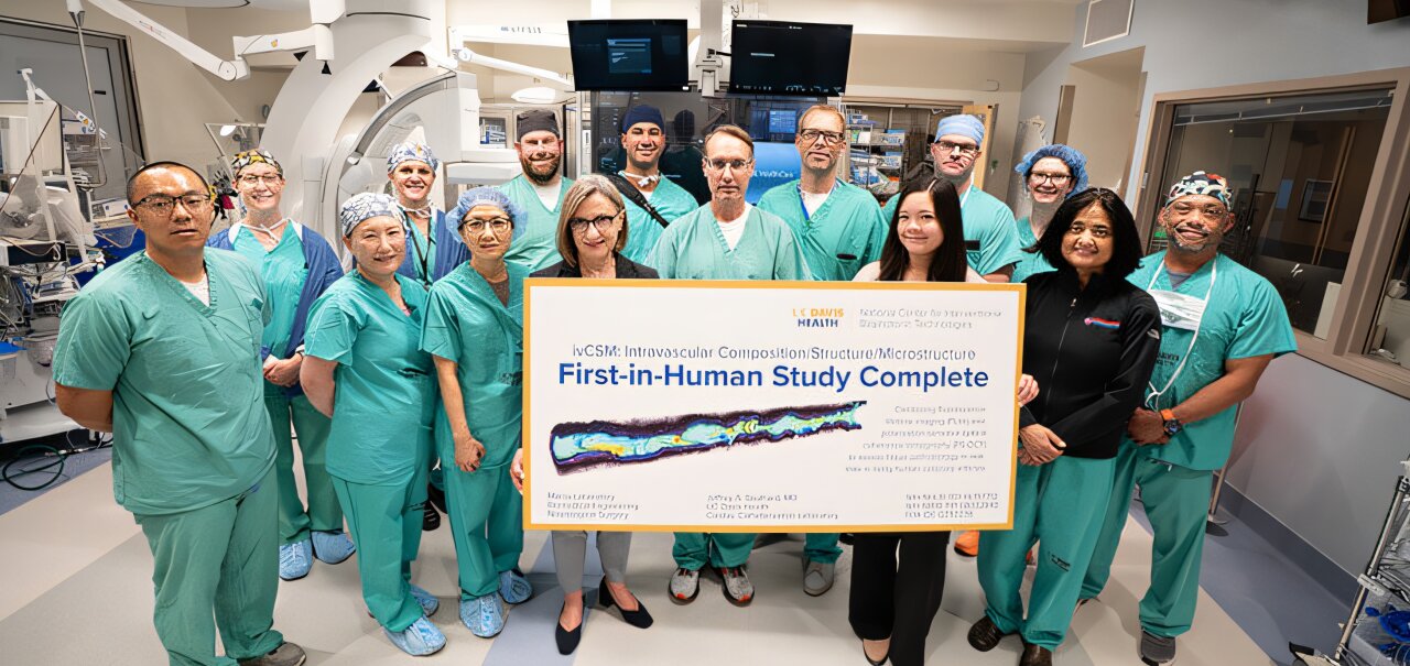
Researchers at the UC Davis National Center for Interventional Biophotonic Technologies (NCIBT) have concluded the first-in-human test of a new device that could help doctors better detect and treat atherosclerosis, the narrowing of blood vessels due to plaque buildup. The flexible catheter-based device, developed at NCIBT labs, is designed to navigate the interior walls of arteries and capture detailed information about plaque buildup in real time.
“Testing this device marks a major step toward advancing heart health. Our technology could help clinicians better understand atherosclerotic cardiovascular disease progression and personalize treatment,” said Laura Marcu, founding director of NCIBT. Marcu holds the Endowed Professorship in Neurosurgical Research and is a professor in the departments of Neurological Surgery and Biomedical Engineering.
The device uses fluorescence lifetime imaging (FLIm) and polarization-sensitive optical coherence tomography (PS-OCT) to simultaneously identify the microstructure and biochemical composition of plaque in coronary arteries.
Julien Bec, who directs engineering and regulatory work in the Marcu Lab, designed and built the catheter and the imaging console.
“FLIm measures the faint natural fluorescence of the artery wall, which reflects its biochemical composition. PS-OCT measures light scattered back from the same tissue to map its microstructure,” Bec explained. “The fact that we are able as an academic group to run this first-in-human study with a catheter system that was developed in a laboratory is unique. This is typically done by big companies.”
The device was previously tested in animal models and in samples of human coronary arteries.
This year, the team received FDA approval to evaluate the technology as an investigational device in 10 patients. In August, they completed imaging for the final patient.
Advancing the detection of heart disease risks
Cardiovascular disease is the leading cause of death in the United States. The buildup of plaque in the arteries can lead to heart attack, stroke and other heart conditions.
Cardiac catheterization (cardiac cath or heart cath) is used to check a heart’s health and diagnose potential heart problems. The doctor inserts a thin, flexible tube into a blood vessel to examine the flow and identify plaque.
Currently, heart cath uses technologies such as optical coherence tomography and intravascular ultrasound to look at the structure and microstructure of blood vessels. It does not show their biochemical composition, which plays an important role in plaque instability and rupture.
The new NCIBT device that combines FLIm with PS-OCT technologies will allow the characterization of both microstructure and biochemistry in a single pullback, a mechanism to withdraw the catheter while taking live images.
“Imagine that you have a very tiny catheter that is compatible to go into the human heart, to rotate, pull back and acquire very fast images that characterize the composition and the microstructure of these blood vessels,” Marcu said.
The catheter goes through the coronary vessels into the beating heart. On its way, it acquires color images that show both the structure and the composition of the artery wall. In two seconds, the device images an 80-millimeter length of a coronary artery as it is pulled back.
The catheter can show plaque buildup even before it develops into stenosis—the abnormal narrowing of blood vessels that can reduce normal blood flow.
“Plaque can rupture suddenly and create blood clots that could lead to a heart attack,” said UC Davis interventional cardiologist Jeffrey Southard. “The catheter will reliably provide information about the biochemistry of the plaque that may indicate inflammation and possible risk of rupture or erosion. Through more research we will learn the best ways to treat this and other types of plaque.”
Southard led the testing of the device in patients, and postdoctoral researcher Xiangnan Zhou operated the system in the cath lab.
Treatment for stenosis caused by plaque buildup often includes placing stents. How and where to place these stents and what drugs to use will affect the long-term success of treatment.
The team’s next step is to scale up the study to include more patients. “We hope that this technology will be used in cath labs for assessing the composition or the pathobiology of plaques in patients and thus inform the treatment and deployment of stents,” Marcu said.
Key medical concepts
Clinical categories

Citation:
First-in-human study of dual-imaging catheter to assess heart attack risk, guide treatment (2026, October 20)
retrieved 20 October 2026
from https://medicalxpress.com/news/2026-10-human-dual-imaging-catheter-heart.html
This document is subject to copyright. Apart from any fair dealing for the purpose of private study or research, no
part may be reproduced without the written permission. The content is provided for information purposes only.